
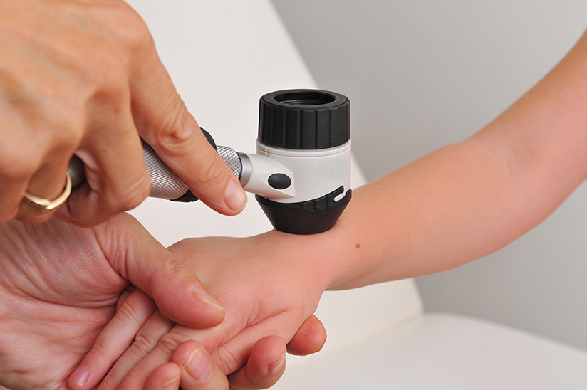
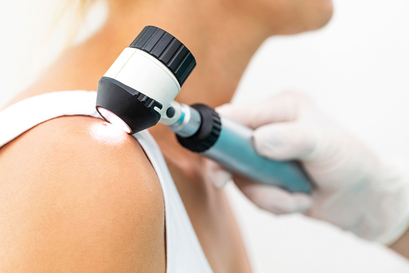
Why does screening for skin cancer work? First of all, through a unique, non-invasive observation device through dermatoscopy, a specialist melanoma surgeon uses you through a full-body skin examination, checking all places which they felt were suspicious. This instrument uses intense light and for further thorough study, expands that picture of the spot.
Stage two used by Visit a Sundoctors in a skin cancer clinic in Melbourne is whole-body imaging, where photographs of every part of their skin are captured. Those images are preserved in such a secure database. When the patient comes back towards an advanced session, the physician examines the samples or images, specifically looking for changes in the various parameters of any suspected moles.
Mole Mapping
When the result for melanoma diagnostic tests, skilled mole monitoring was provided inside a mole monitoring hospital. Complete body mole mapping is a method of tracking the entire surface of the body and particular moles or lesions’ locations. A body mole chart enables present and past images of moles on the body to be matched with pictures taken in the future. The best strategy to track the emergence of new moles is to have a mole road map made.
The Difference Between Mole Mapping And Skin Cancer Check
A mole map effectively takes a series of 28 images covering the body’s outer structure. These are then placed in a database where the comparison to previous mole map images could be correlated with them.
Skin cancer management requires specialist skin cancer doctors who conduct dermatoscopic examinations of the skin from head to toe. If they show any symptoms of melanoma, certain moles and freckles will be carefully analyzed and tested.
How Does Mole Mapping Work?
Mole mapping utilizes specialized tools for digital mole mapping –
The mole map approach is non-invasive in itself. A human is a query to take one’s clothes off. The method of getting photos of each and every area of the body then starts. As the images are taken, the operator needs to pause and verify them to ensure that the precision, focus, and positions are right.
Its most advanced optical mole mapping tools and optics accessible on the market are used at the mole screening clinic. To make sure that the entire skin surface is shielded, this technique splits the tissue into 28 parts.
In addition to storing technology images, inbuilt resources are provided that greatly aid in the diagnosis of various forms of precancerous moles and skin cancer. The ability to detect good locations or lesions is among the most significant features of mole mapping technology.
New software and optics advances and the ability to store vast volumes of data have rendered mole mapping an effective diagnostic method for skin cancer diagnosis.
Conclusion:-
Australia has the world’s highest levels of skin cancer, making early detection of melanoma important. Without showing any symptoms or apparent indications of massive change, melanoma and other skin cancer grow more slowly. At the earliest possible time, mapping significantly increases the chances of detection.